About our software
CCMetrics and ACCMetrics are validated image analysis software for manual and automated tracing of corneal nerve morphology.
The software can be used for three main CCM measures:
- Corneal nerve fibre density (CNFD) (fibres/mm2)
- Corneal nerve branch density (CNBD) (branches/mm2)
- Corneal nerve fibre length (CNFL) (mm/mm2)
ACCMetrics also quantifies additional variables like the fractals, nerve fibre area, width and total branch density.
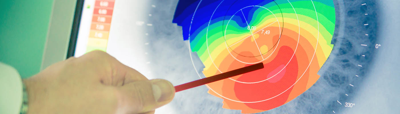
We also offer Sudometrics, an image analysis software that quantifies the percentage colour change in pink over the whole area of Neuropad. Neuropad is a visual screening test that provides a visual means of identifying diabetic patients at risk of foot ulceration.
More about the products
This software has been developed from research carried out by the Early Neuropathy Assessment (ENA) Group at Weill Cornell Medicine – Qatar and The University of Manchester.
Further details about each product can be found on the relevant license agreement page.
CCMetrics
Corneal nerve fibre quantification.
ACCMetrics
Corneal nerve fibre analyser.
Sudometrics
Quantification of the colour change of the Neuropad response.
Access our software
We offer our CCMetrics, ACCMetrics and Sudometrics software free of charge for research purposes.
License agreements
You must read the license agreement and complete an online form for the software that you wish to access. You will also find further details of each product at these links:
Training workshops
We also run a CCM workshop for researchers who analyse corneal nerve morphology using the Heidelberg Retina Tomograph 3 device.
Workshops and training videos
Our CCM workshop is for researchers who analyse corneal nerve morphology using the Heidelberg Retina Tomograph 3 device.
Using our established Manchester protocol, you will be trained on how to acquire and extract corneal nerve images. You will be taught how to analyse corneal nerve morphology using CCMetrics and the ACCMetrics for manual and automated tracing of the nerve fibres.
The workshop runs over three to five days, depending on your skills and background.
This workshop is offered by the ENA Group for free. You can attend this workshop either at Weill Cornell Medicine in Qatar or at The University of Manchester in the UK.
You are not required to undertake the workshop before you can access the CCMetrics and ACCMetrics software, but it may be useful for those who need to learn about the technique and image analysis.
Please contact us for more information about the workshop.
Training videos
You may also find the following training videos on CCM assessment useful:
- Part 1 – Performing CCM assessment and image acquisition (YouTube)
- Part 2 – Extracting CCM images (YouTube)
- Part 3 – CCM image analysis (YouTube)
Citing our software and research
If you publish results using CCMetrics/ACCMetrics, please mention the software in the methods section of your paper. Acknowledgement of the ENA Group is always appreciated.
Here is a list of our papers on the design and validation of the CCMetrics and ACCMetrics, should you wish to cite them in your paper:
- Normative values for corneal nerve morphology assessed using corneal confocal microscopy: a multinational normative data set. Tavakoli M et al. Diabetes Care. 2015; 38: 838-43.
- Rapid automated diagnosis of diabetic peripheral neuropathy with in vivo corneal confocal microscopy. Petropoulos I N, et al. Invest Ophthalmol Vis Sci. 2014; 55: 2071-8.
- Small nerve fiber quantification in the diagnosis of diabetic sensorimotor polyneuropathy: comparing corneal confocal microscopy with intraepidermal nerve fiber density. Chen X et al. A. Diabetes Care. 2015; 38: 1138-44.
- Diagnostic utility of corneal confocal microscopy and intra-epidermal nerve fibre density in diabetic neuropathy. Alam U, et al. PLoS One. 2017 Jul 18;12(7).
- Corneal confocal microscopy is a rapid reproducible ophthalmic technique for quantifying corneal nerve abnormalities. Kalteniece A et al. PLoS One. 2017 Aug 17;12(8): e0183040. doi: 10.1371/journal.pone.0183040. eCollection 2017.
- Diagnosis of Neuropathy and Risk Factors for Corneal Nerve Loss in Type 1 and Type 2 Diabetes: A Corneal Confocal Microscopy Study. Ferdousi M et al. Diabetes Care. 2021 Jan;44(1):150-156. doi: 10.2337/dc20-1482. Epub 2020 Nov 3. PMID: 33144353; PMCID: PMC7783929.
- The Inferior Whorl For Detecting Diabetic Peripheral Neuropathy Using Corneal Confocal Microscopy. Petropoulos IN et al. Invest Ophthalmol Vis Sci. 2015 Apr;56(4):2498-504. doi: 10.1167/iovs.14-15919. PMID: 25783609; PMCID: PMC4408884.
Contact us
If you have any questions about our software or the ENA Group, please get in touch.