Delayed reperfusion deficits after experimental stroke account for increased pathophysiology.
Burrows FE, Bray N, Denes A, Allan SM, Schiessl I. (2014) “Delayed reperfusion deficits after experimental stroke account for increased pathophysiology.” J Cereb Blood Flow Metab Nov 19.
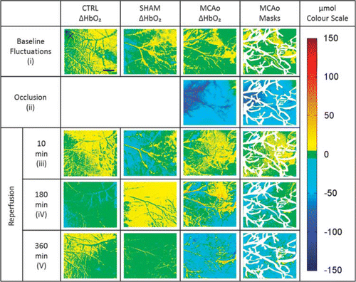
Cerebral blood flow and oxygenation in the first few hours after reperfusion following ischemic stroke are critical for therapeutic interventions but are not well understood. We investigate changes in oxyhemoglobin (HbO2) concentration in the cortex during and after ischemic stroke, using multispectral optical imaging in anesthetized mice, a remote filament to induce either 30 minute middle cerebral artery occlusion (MCAo), sham surgery or anesthesia alone. Immunohistochemistry establishes cortical injury and correlates the severity of damage with the change of oxygen perfusion. All groups were imaged for 6 hours after MCAo or sham surgery. Oxygenation maps were calculated using a pathlength scaling algorithm. The MCAo group shows a significant drop in HbO2 during occlusion and an initial increase after reperfusion. Over the subsequent 6 hours HbO2 concentrations decline to levels below those observed during stroke. Platelets, activated microglia, interleukin-1α, evidence of BBB breakdown and neuronal stress increase within the stroked hemisphere and correlate with the severity of the delayed reperfusion deficit but not with the ΔHbO2 during stroke. Despite initial restoration of HbO2 after 30 min MCAo there is a delayed compromise that coincides with inflammation and could be a target for improved stroke outcome after thrombolysis.
Read the full article here.


Contact Us
+44 (0) 161 306 6000
0 Comments