The Difficulties of Communicating What You Do (Not) See: Pamela Mackenzie on Early Modern Plants, Microscopy, and Visual Knowledge
Stefan Hanß: The support of early career researchers is a strong element of the upcoming British Academy event Microscopic Records: The New Interdisciplinarity of Early Modern Studies, c.1400–1800. One of the speakers will be Pamela Mackenzie, a PhD candidate at the University of British Columbia’s Department of Art History, Visual Art and Theory. She is currently a predoctoral researcher in Dr Sietske Fransen’s Max Planck research group Visualizing Science in Media Revolutions at the Bibliotheca Hertziana/Max Planck Institute for Art History in Rome. Pamela is interested in the relationship between early modern prints and visual epistemologies, thus, processes of knowing that take place via images and visual communication more broadly speaking. Her PhD thesis explores some of the earliest images produced of plants as seen through microscopes. In the seventeenth century, when the microscope had been a newly developed technology, natural historians and botanists like Nehemiah Grew pioneered the exploration of microscopic records of the botanical world (fig. 1). What fascinates you of these images and how did you end up working on the history of early modern microscopy?
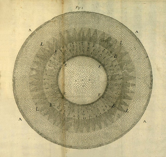
Fig. 1: Nehemiah Grew, The Comparative Anatomy of Trunks (London, 1675), “A slice of the lower part of the Root of Horse-Radish cut traversly,” engraving. Image credit: The Public Domain Review, https://publicdomainreview.org/collection/nehemiah-grew-s-anatomy-of-plants-1680.
Pamela Mackenzie: I am an art historian by training but throughout the course of this degree I’ve become more and more interested in the history of science. I’ve been finding that a lot of the visual analysis skills that I developed by spending so much time working directly with images have given me a lot to contribute to early modern history of science. My work draws connections between knowledge production and illustration through an analysis of early modern prints. In other words, I’m interested in the way that illustrations allowed people to communicate what they had seen to an audience of others who might not have been able to see the same things. Maybe that was because they lacked the ‘right’ tools—say, for example, Galileo’s descriptions of Jupiter’s moons that he was only able to see because of his telescope, or because the objects were far away like all the drawings of plant species from the Americas that Europeans sent back overseas. In these cases, the illustrations were able to show the existence and properties of objects that would have otherwise been completely inaccessible for many who saw those images.
I am convinced that spending a lot of time with an image and taking it really seriously as an artifact that is neither merely aesthetic nor instructional can bring out a lot of meaning that gets lost easily when illustrations are treated more as decorations or afterthoughts to the development of the scientific process. Images that are immediately striking or visually interesting, like the above illustration of a heavily magnified horseradish root (fig. 1), contain a great deal of technical information and were produced after prolonged, expert engagement with the subject matter. These images would have been examined closely by the authors’ and artists’ peers and were crucial for transmitting information and new theories about how the natural world operates. The fact that early scientific illustrations also tended to be beautiful was by no means incidental—even today we are regularly confronted by stunning, high-definition renderings of data and astronomical phenomena beyond the reaches of the naked eye—but these images also served an important role in shaping and sharing knowledge.
SH: I would be curious to know whether there are particular images that arouse your interest in the first place.
PM: An especially exciting area of visual production for me has been in the images produced by early users of microscopes in the late seventeenth century. The most famous examples of these sorts of images are Robert Hooke’s magnificent illustrations that accompanied his seminal text on microscopy, Micrographia, published by the Royal Society London in 1665. Particularly well-known is a large, fold-out illustration of a flea that features exquisite line work and meticulous detailing, the sense of scale made apparent in the individual rendering of hairs across the back and legs of the flea (fig. 2). I like to imagine that I am sharing a sense of wonder at both the beauty that can be found in the small details of nature through close looking, and also at the skill of the artist and early scientific practitioner Robert Hooke (1635–1703).
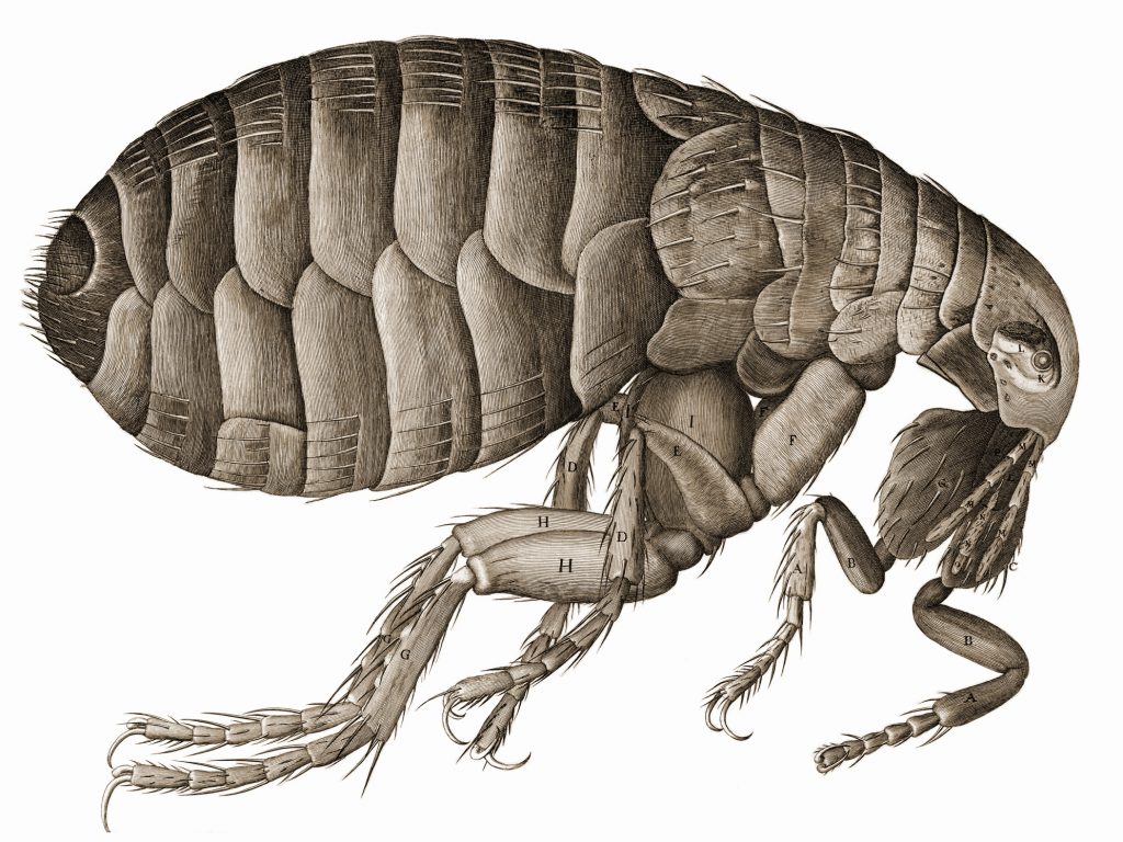
Fig. 2: Robert Hooke, Micrographia (London, 1665), engraving of a flea. Image credit: Wikimedia commons/The National Library of Wales, https://en.wikipedia.org/wiki/File:HookeFlea01.jpg.
SH: An earlier blog entry explores such experiences associated with early modern microscopic records by means of visual de- and recontextualisation. Can you elaborate a bit more on the work by Robert Hooke, and how it does relate to your PhD research?
PM: My dissertation focuses on the work of one of Robert Hooke’s peers at the Royal Society, Nehemiah Grew. Working in the decades after Hooke, Grew turned the lens of the microscope to the study of plants. Using his training as a physician, Grew carefully dissected trees, seeds and flowers in order to explore their mechanisms of growth and reproduction. One of his most famous work is the Anatomy of Plants (London, 1682). The images that accompanied Grew’s publication on plant anatomy were truly mesmerizing. His often abstract and geometric interpretation of plant life at a microscopic scale, intricately patterned with circles and hatching, can be disorienting until one realizes that the subject matter is intended to be heavily magnified cross-sections of plants (fig. 3). What we can learn by analyzing these images closely and reading the corresponding texts is that Grew was not just making images of what he saw directly through the microscope. Of course, these images do correspond to actual specimens that he saw, but his choice to represent these plants with such rigid and regular forms also reflects his interest in uncovering underlying principles in nature: he believed that plants grew in fixed patterns according to geometrical principles. While this doesn’t exactly agree with contemporary scientific understandings of plant growth, it does tell us a lot about how some seventeenth-century observers were approaching their studies of nature.
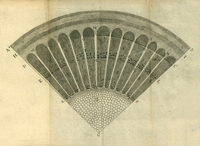
Fig. 3: Nehemiah Grew, The Comparative Anatomy of Trunks (London, 1675), “a quarter of a slice of a Branch of Barbery Tree of 2 years growth”, engraving. Image credits: The Public Domain Review, https://publicdomainreview.org/collection/nehemiah-grew-s-anatomy-of-plants-1680.
SH: …and how they wished the results of their studies be seen by others. Could you say a bit more on how you wish others to see your own research? What is your work contributing to the history of microscopy?
PM: What we have to keep in mind as modern readers and viewers of these images is that what was being seen by individuals like Hooke and Grew had effectively never been seen before. They were interpreting microscopic structures for the first time and trying to make sense of what they saw in order to communicate that knowledge to a community of their peers. The illustrations were a way for others, who either did not have access to a microscope or who were occupied with their own microscopic studies, to see what they were seeing. The images were intended to encode and transmit information about previously unseen microscopic dimensions of the natural world to a visually literate and educated audience. My work, today, involves being attentive to the details included in these illustrations, to pay attention to the choices that were made when creating representations of nature, in order to better understand what was important to early scientific communities in the seventeenth century.
SH: Thank you! We are excited to hear more about that at the upcoming British Academy event!
Further reading:
For anyone interested in browsing more illustrations made by early members of the Royal Society, we recommend the Royal Society’s new picture library. Using the search tool is a great way to find images in areas of early scientific study that may be interesting for you: for example, the term ‘microscope’ turns up many fascinating illustrations of diverse subjects like bug eyes and magnified crystals.
Pamela Mackenzie, University of British Columbia/Bibliotheca Hertziana – Max Planck Institute for Art History
Stefan Hanß, The University of Manchester


Contact Us
+44 (0) 161 306 6000
0 Comments